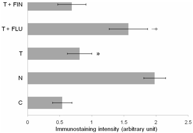
Figure 5.

Quantitative analysis of MECA-79 immunoperoxidase staining intensity at the apical membrane of the luminal epithelia in different experimental groups. The highest intensity was observed in rats received normal steroid replacement regime and following co-administration of flutamide with testosterone during uterine receptivity period. Mean was obtained from intensity of 4 different sections per treatment group. *P < 0.05 as compared to normal steroid replacement, †P < 0.05 as compared with testosterone only treatment. C: vehicle control, N: normal steroid replacement regime, T: testosterone only injection with P4, T + FLU: testosterone and flutamide administration with P4 and T + FIN: testosterone and finasteride administration with P4.
