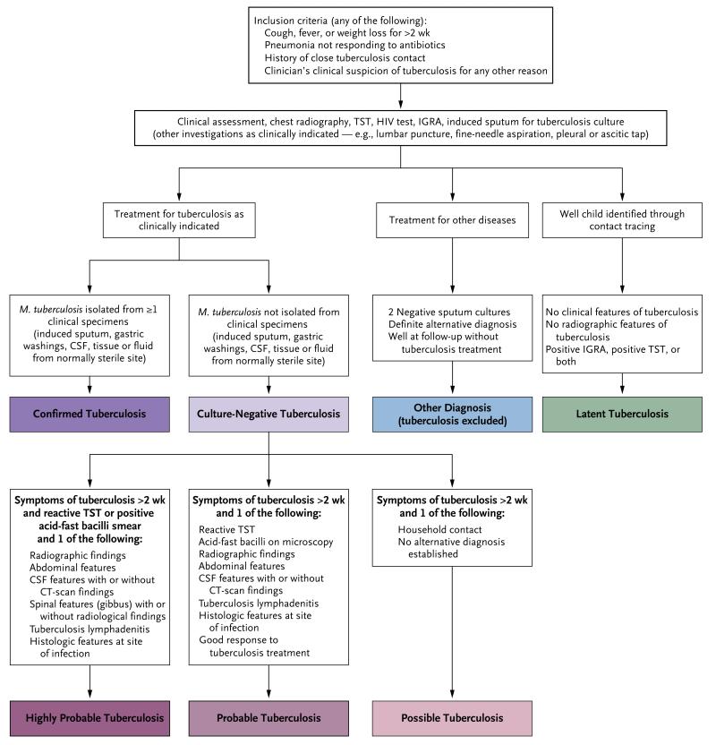
Figure 2. Diagnostic Algorithm.
With regard to the inclusion criteria, patients with failure to thrive for more than 4 weeks were included in the Kenyan cohort. In the group receiving treatment for other diseases, patients with IGRA-positive results were excluded from the South Africa and Malawi cohorts but included in the Kenyan cohort. In the culture-negative group, the IGRA was repeated for patients in whom tuberculosis was suspected and the initial IGRA was negative; findings on radiography included effusion, extensive consolidation, cavitation, lymphadenopathy, miliary disease, and lobar pneumonia that was not responding to antibiotics, and abdominal features included ascites and lymphadenopathy. CSF denotes cerebrospinal fluid, CT computed tomography, IGRA interferon-γ-release assay, and TST tuberculin skin test.

