
Fig. 1.
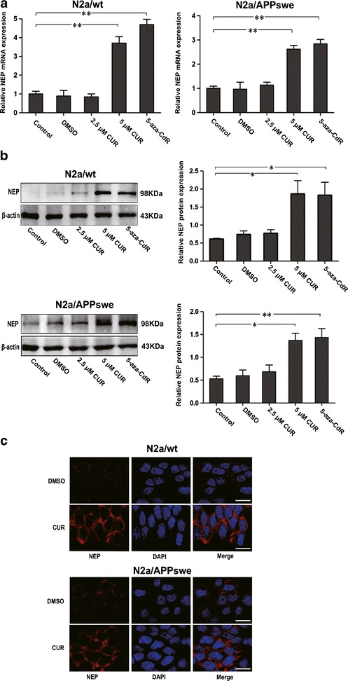
Effects of CUR treatment on NEP expression in N2a/wt and N2a/APPswe cells. N2a cells were incubated with DMSO (0.1%), CUR (2.5 μM, 5 μM) for 48 h, or 5-aza-CdR (5 μM) for 72 h. Cells in each group were then collected; RNA and proteins were assessed. a qPCR analysis of NEP mRNA level. GAPDH was assessed as a loading control. b Western blot analysis of NEP protein level. β-actin was assessed as a loading control. c Immunocytochemistry analysis of NEP protein expression and distribution. N2a cells were treated with 5 μM CUR for 48 h and labeled with anti-NEP antibody (red) and DAPI (blue). Scale bar 100 μm. All data were represented as a mean ± SD of three independent experiments. *p < 0.05, **p < 0.01