
Figure 4.
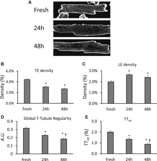
AutoTT analysis of T-tubule remodeling of adult murine ventricular cardiomyocytes in culture. (A) Examples of confocal images of freshly isolated myocytes and myocytes cultured for 24 or 48 h. Images were subjected to AutoTT analysis for TE (B) and LE (C) density, regularity (D), and integrity of Global T-tubules (TTint, E). ∗p < 0.05 vs. freshly isolated myocytes; †p < 0.01 vs. 24 h in culture. n = 36, 38, and 38 cells for fresh, 24 h in culture and 48 h in culture, respectively.