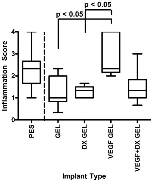
Figure 7.
Inflammation score of tissues surrounding implants after 2 weeks implantation in rat subcutaneous tissue. The implanted materials were bare PES fibers (PES, n = 9), hydrogel-coated PES fibers (GEL, n = 9), VEGF-releasing hydrogel-coated PES fibers (VEGF GEL, n = 6), DX-releasing hydrogel-coated PES fibers (DX GEL, n = 8), and VEGF + DX-releasing hydrogel-coated PES fibers (VEGF + DX GEL, n = 9). The tissues surrounding VEGF-releasing hydrogel-coated fibers were significantly inflamed compared to tissue surrounding hydrogel-coated and DX-releasing hydrogel-coated fibers (p < 0.05).

