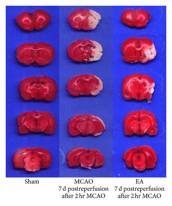
Figure 2.

TTC staining of brain slices. Ischemic area is white; intact area stained red. Representative coronal sections of sham group, MCAO groups at 2 h MCAO followed by 7 d of reperfusion, and the EA groups of 2 h MCAO followed by 7 d of reperfusion.
