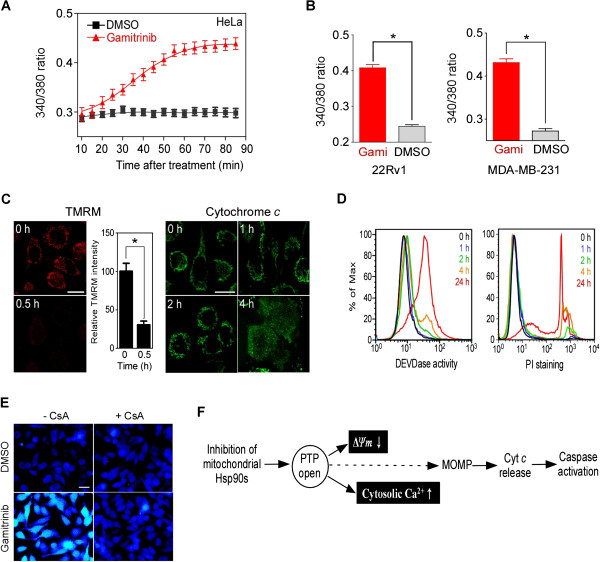
Figure 1.

Mitochondrial Hsp90s modulate the mitochondrial calcium store. (A) Time course of cytosolic calcium increase. The ratio of the emission fluorescence intensities at 340 and 380 nm excitation of Fura-2 labeled HeLa cells in calcium-free medium was measured after 30 μM gamitrinib treatment as described in Materials and Methods. (B) Increase of cytosolic calcium in 22Rv1 and MDA-MB-231 cells. Fura-2 fluorescence ratio after 30 μM gamitrinib (Gami) treatment for 1 hour was calculated. Data are the mean ± SEM of duplicated experiments and collected from 40 regions of interest (ROIs). (C) Mitochondrial membrane permeabilization. TMRM-loaded HeLa cells were imaged to measure mitochondrial membrane potential depolarization (ΔΨm) (left); alternatively, cytochrome c redistribution was analyzed (right) at the indicated times after 30 μM gamitrinib treatment as previously described [35]. White bar, 20 μm. (D) Caspase activation and cell death induction. After 30 μM gamitrinib treatment, HeLa cells were labeled with FITC-DEVD-fmk (left, DEVDase activity) or propidium iodide (right, PI staining) and analyzed by flow cytometry at the selected time points. (E) Cyclosporin A (CsA) blocks cytosolic calcium increase. Cytosolic calcium changes in Fura-2-labeled HeLa cells treated for 1 hour with 5 μM CsA and/or 30 μM gamitrinib were analyzed. Bar, 50 μm. (F) Summary of sequential events following mitochondrial Hsp90 inhibition. PTP opening is directly linked with the loss of ΔΨm and increase of cytosolic calcium. The calcium flux occurs prior to mitochondrial outer membrane permeabilization (MOMP) and cytochrome c release. *, p < 0.0001.
