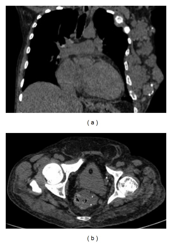
Figure 3.

A 60-year-old man with Klippel-Trenaunay-Weber syndrome presenting asymmetric growth of the lower limbs. CT of the chest showed increased soft tissue as well as extensive vascular malformations in the left hemithorax wall with intermingled phleboliths, causing multiple lytic lesions with enlargement in the ipsilateral ribs (a). CT of the abdomen showed a thick-walled rectum intermingled with phleboliths, denoting varicose veins (b).
