
Figure 2.
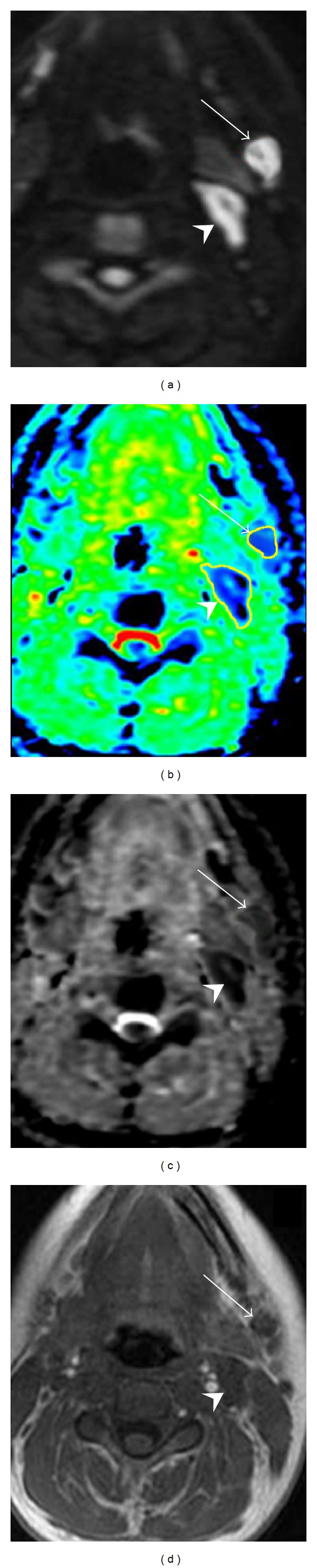
Axial native DWI image at b value of 1000 sec/mm2 (a), color ADC map (b), grey scale ADC map (c), and T1-weighted image (d) of a patient with nasopharynx carcinoma showing two solid lymphadenopathies located at level Ib (arrow) and level IIa (head arrow), respectively. According to morphological criteria the lymph node at level Ib, round shaped and 6 mm in size, was considered to be negative for cancer, while the adenopathy localized at level IIa, oval shaped and with a short transverse diameter of 11 mm, was reported to be suspicious for metastatic involvement. Concerning DWI, level Ib lymph node showed a mean ADC value of 0.690 × 10⁻³ mm2/sec and was considered suspicious for metastatic involvement, whereas level IIa adenopathy displayed an average ADC value of 1.051 × 10⁻³ mm2/sec and, therefore, was deemed to be a noncancerous lymphadenopathy. At pathological examination level Ib lymph node showed large intranodal metastatic deposits and level IIa adenopathy was found to be benign.