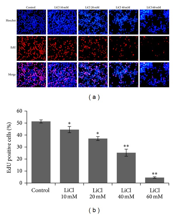
Figure 2.

LiCl inhibited proliferation in SW480 cells. Cell proliferation assay was preformed, in which EdU-labeled proliferative cells (red) and Hoechst-stained nuclei (blue) were observed under a fluorescent microscope. Cells were treated with vehicles (PBS) and different concentrations of LiCl (10 mM, 20 mM, 40 mM, and 60 mM), respectively. Data are representative of at least three independent experiments and are expressed as the mean ± SEM. Original magnification, ×100. *P < 0.05 and **P < 0.01 versus Control group.
