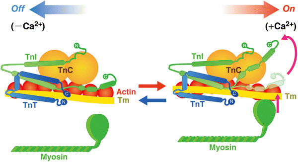
Fig. 3.

Structure and arrangement of cardiac thin filament proteins in the absence (off) and presence (on) of Ca2+. Shapes of the proteins are drawn primarily based on evidence from the work by Takeda et al. [15]. Upon Ca2+ binding to TnC, the C-terminus region of TnI dissociates from actin, allowing for Tm movement and, consequently, myosin binding to actin. Arrows indicate relative movements of Tm position in diastole and systole. C COOH terminus, N NH2 terminus. The equilibrium between the “off” state and the “on” state is a function of [Ca2+]i
