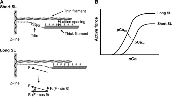
Fig. 5.

Schematic illustration showing the role of titin in the modulation of interfilament lattice spacing (a) and length-dependent changes in the force–pCa curve (b). At a long length, titin produces passive force (F) in the myofilament lattice. The force is divided into longitudinal (F L) and radial (F R) components, the latter of which reduces interfilament lattice spacing. The lattice reduction is likely to enhance myosin attachment to actin and, consequently, it causes a leftward shift of the force–pCa curve and increases the maximal Ca2+-activated force [see arrow (indicating the mid-point of the curve) in (b)]
