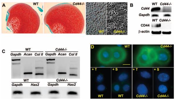
Figure 1.
Characterization of cartilage and primary chondrocytes from BALB/c wild-type (WT) mice and CD44−/− mice. A, Femoral heads isolated from WT and CD44−/− mice were fixed directly and stained with Safranin O–fast green (top). Primary murine chondrocytes isolated from the femoral heads of WT and CD44−/− mice by collagenase digestion, were plated at high density and observed under phase-contrast microscopy (bottom). B, Chondrocytes from WT and CD44−/− mice were characterized for the expression of CD44 by conventional reverse transcription–polymerase chain reaction (RT-PCR) using GAPDH as control or by Western blotting using an anti-mouse CD44 antibody, with β-actin antibody as control. C, The expression of mRNA for aggrecan (Acan), type II collagen (Col II), and hyaluronan synthase 2 (HAS-2) derived from chondrocytes obtained from WT and CD44−/− mice was determined by conventional RT-PCR using GAPDH as control. Results are representative of 2 independent experiments. D, Chondrocytes from WT and CD44−/− mice were stained for hyaluronan (green) either directly or following pretreatment with testicular hyaluronidase (+T) or Streptomyces hyaluronidase (+S). Results are representative of 5 experiments.

