Figure 3.
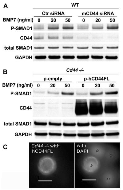
Altered levels of CD44 expression and sensitivity to bone morphogenetic protein 7 (BMP-7). A and B, To examine the effects of a loss of function in relation to CD44, chondrocytes from wild-type (WT) mice were transfected with either control small interfering RNA (siRNA) or mouse-specific CD44 (mCD44) siRNA (A). To examine the effects of a gain of function in relation to CD44, chondrocytes from CD44−/− mice were transfected with a control empty vector (p-empty) or with cDNA encoding full-length human CD44H (p-hCD44FL) (B). Two days after transfection, the cells were stimulated with 0, 20, or 50 ng/ml of BMP-7 and then analyzed by Western blotting for changes in Smad1 phosphorylation (p-Smad1). CD44 protein expression was probed with an anti–CD44 cytotail antibody. Total Smad1 and GAPDH were used as loading controls for protein content. Each blot is representative of 3 replicate experiments. C, Human CD44FL was expressed at the surface of chondrocytes from CD44−/− mice transfected with hCD44FL (left). Overlay of DAPI staining of nuclei of the same image is also shown (right). Asterisks identify nuclei of cells within the same field. Image is representative of 2 separate experiments. Transduction with a control empty vector yielded no immunofluorescence for human CD44 (data not shown).

