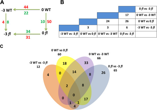
Figure 4.

Summary of differentially phosphorylated proteins identified in this study. A: Number of phosphoproteins differentially expressed during fiber development within and between WT and fl ovules. Numbers above and below arrows denote numbers of phosphoproteins differentially expressed for the specified comparison. For example, between stages -3 and 0 DPA in the WT, 44 phosphoproteins were up-regulated (red) and 22 were down-regulated (blue) at 0 DPA. Similarly, between WT and fl at -3 DPA, 4 phosphoproteins were up-regulated and 8 phosphoproteins were down-regulated in the WT. B: Numbers at column-row intersections are the number of differentially phosphorylated proteins common to the four tissues. C: Venn diagram showing the number of differentially phosphorylated proteins shared between phosphoproteomes of the different four tissues.
