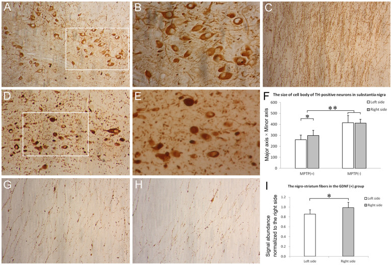
Figure 6. Immunohistochemical analysis of dopaminergic system.
Compared with naive controls (A and B), the number of TH-positive neurons in the substantia nigra pars compacta was dramatically reduced in all MPTP-treated monkeys (D and E). (B) and (E) represent the magnified views of the insets in (A) and (D), respectively. However, no significant difference existed in the number of TH+ neurons in the left versus right half of substantia nigra either in the GDNF (+) or GDNF (-) group after MPTP treatment. The signal abundance of TH immunoreactivity on the nigro-striatum fibers was also reduced after MPTP treatment (C versus G and H). (F) The size of the remaining TH-positive neurons (minor axis equal or more than 10 μm) in substantia nigra was quantified. The neurons were smaller after MPTP treatment and displayed slight but significant difference in size in the right versus left half of substantia nigra. (I) The signal abundance of the nigro-striatum fibers was quantified within the GDNF(+) group. (A), (D) and (C), 200 ×; (B), (E), (G) and (H), 400 ×. *P < 0.05, **P < 0.01.

