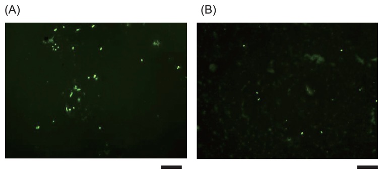
Fig. 2.
Fluorescent microscopic image of microbes collected with an adhesive sheet. Microbial cells were stained with 1×SYBR Green II (scale bars, 10 μm). (A) Mixture of A. lwoffii ATCC15390, B. subtilis 168, P. putida ATCC12633 and S. epidermidis IFO3762; (B) sample from vertical surface of the rack in our laboratory.

