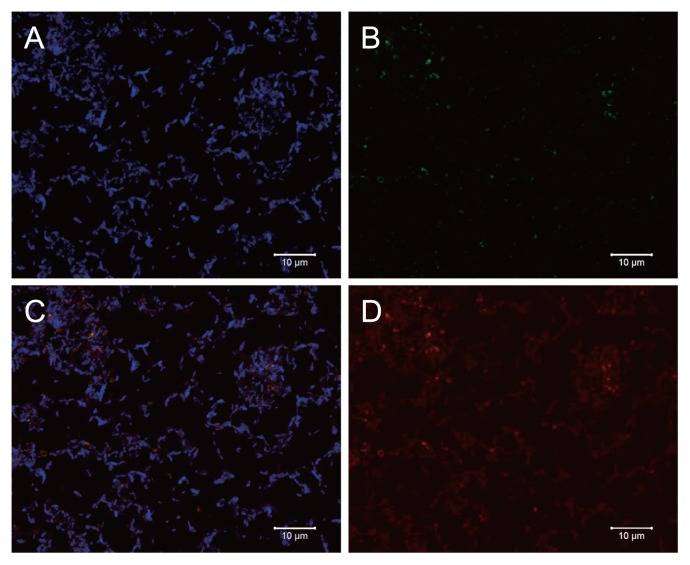
Fig. 3.
Epifluorescence micrographs of in situ hybridization of the enrichment archaeon Kjm51a grown on MYB medium for a week. The same microscopic field is shown after hybridization with a Kjm51a-specific probe (red), an archaeal probe ARC915 (green), and a bacterial probe EUB338 (blue). A, blue color; B, green color; C, merge of red, green and blue colors; D, red color. Bars, 10 μm.

