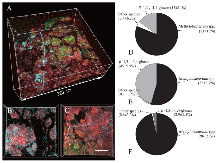
Fig. 3.
FISH assay and microbial community composition of pink biofilms. A–C, CLSM photomicrographs showing the spatial distribution of microorganisms in three independent pink biofilms. The organisms were targeted by in situ hybridization with the ROX-labeled probe MB and FITC-labeled probe EUB338, and simultaneously stained with calcofluor white. Cells of MB-stained Methylobacterium are red; cells of EUB338-stained bacteria are green. Cells containing β-1,3-glucan like fungi are aqua blue. D–F, pie charts of Methylobacterium, other bacteria, and β-1,3-, 1,4-glucans. Methylobacterium is the bacterial group that hybridized with MB, other bacteria are the bacterial group that hybridized with EUB338, and β(1–3) and β(1–4)-linked glucosyl polymers are the positions that hybridized with calcofluor white. Values are the mean ± standard deviation for duplicate samples.

