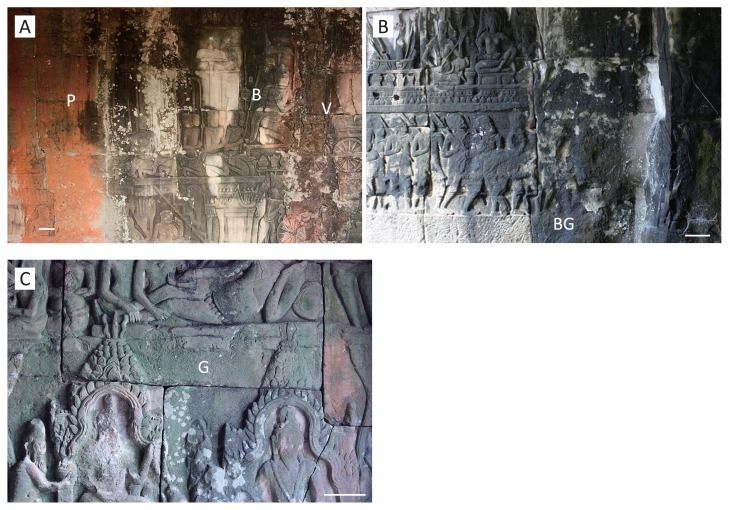
Fig. 2.
Various pigmented biofilms formed on the sandstone walls of the inner gallery of Bayon. Bar indicates 10 cm.
A. Salmon pink (P), black–gray (B), and signal violet (V) biofilms on the north-facing wall on the north side. Samples P53, B53, and V53 were obtained from the positions indicated “P”, “B”, and “V”, respectively.
B. Blue–green (BG) biofilm on the east-facing wall on the south side. Samples BG1, BG4, and BG5 were obtained from the position indicated “BG”, measuring 30 cm in circumference. Sample BG4 was used for analysis of bacterial stratified structure.
C. Chrome green (G) biofilm on the south-facing wall of the east side. Sample G27 was obtained from the position indicated by “G”.

