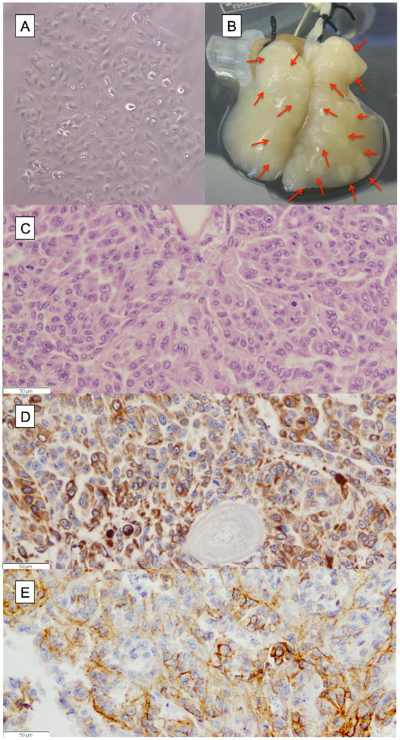
Figure 1.

A549 cells grown on tissue of the 4D model and the 2D flask. (A) A549 cells growing in a culture flask in 2D fashion. (B) Photograph of tissue of the 4D model with multiple tumor nodules (red arrows). (C) The H&E stain (40X) showing cell-cell and cell-matrix interaction of cells growing in the 4D model supported by IHC staining of the cells with CK7 (D) and E-cadherin (E).
