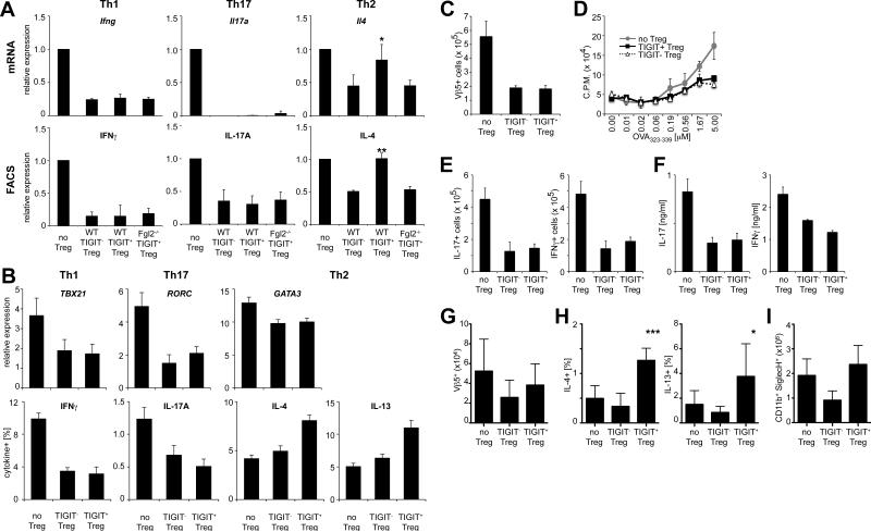
Figure 5. TIGIT+ Treg cells suppress Th1 and Th17 cell but not Th2 cell responses.
(A) Naïve effector T cells, WT Foxp3+TIGIT−, WT Foxp3+TIGIT+ Treg cells, and Fgl2−/− Foxp3+TIGIT+ Treg cells were sorted and co-cultured at a ratio of 1:10 under Th1, Th2, or Th17 polarizing conditions. After 3 days mRNA was measured by quantitative RTPCR. On day 5 intracellular cytokines in CD45.1+ T effector cells were determined by flow cytometry (values normalized to unsuppressed controls, mean ± SEM; * P<0.05, ** P<0.001, paired student's t-test). (B) Human TIGIT+ and TIGIT− Treg cells (CD4+CD25highCD127neg) were sorted and co-cultured with CFSE-labeled CD25-depleted CD4+ T effector cells. Gene expression (qRT-PCR) and intracellular cytokines (flow cytometry) were measured on day 4 (mean ± SEM; n=6). (C-F) CD25− effector OT-II cells and CD25high OT-II Treg cells (TIGIT−, TIGIT+ or no Treg cell control) were transferred i.v. into WT recipients and mice were immunized with OVA in CFA. (C) Expansion of Vβ5+ OT-II T cells and (D) proliferation in response to OVA323-339 were determined 10 days later. (E) Intracellular cytokine levels were determined by flow cytometry and (F) cytokine concentration in the culture supernatants was determined by cytometric bead array. (G-I) CD25− effector OT-II cells and CD25high OT-II Treg cells (TIGIT−, TIGIT+ or no Treg cell control) were transferred i.v. into WT recipients. Mice were then sensitized with OVA (i.p.) on days 0 and 7 and challenged with aerosolized OVA on days 14–17 to induce allergic airway inflammation. (G) Total numbers of Vβ5+ OT-II cells in lungs, (H) intracellular cytokine levels from lung-infiltrating CD4+ T cells, and (I) total eosinophil numbers in bronchio-alveolar lavage fluid were determined by flow cytometry. Pooled data from two experiments are shown (mean ± SEM; n=8).

