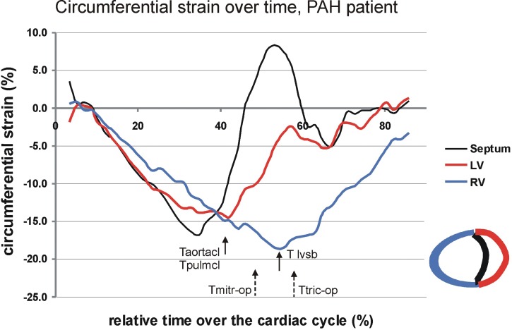
Figure 1.
The presence of ventricular dyssynchrony illustrated in a patient with pulmonary arterial hypertension (PAH) measured by cardiac magnetic resonance imaging (CMR) strain analysis. Circumferential strain curves reflect myocardial shortening (negative strain) and stretching (positive stain) over time of the cardiac cycle for the right ventricular (RV) free wall (blue), left ventricular (LV) free wall (red), and interventricular septum (black). The LV, RV, and septum start simultaneously with shortening. However, RV peak shortening occurs later than LV peak shortening in the cardiac cycle. The aortic and pulmonary valves close (Taortacl and Tpulmcl) at the time of LV peak shortening. The time of maximal leftward septal bowing (Tlvsb) occurs coincidently with septal stretching and with peak shortening of the RV. Tmitr-op and Ttric-op indicate the opening times of the mitral and tricuspid valves and indicate the onset of LV and RV filling.

