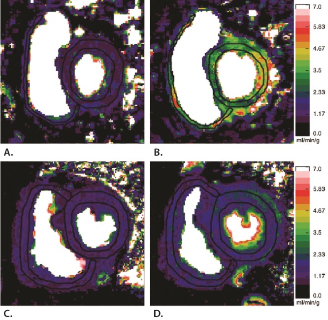
Figure 3.

Representative perfusion maps showing that biventricular perfusion reserve was diminished in patients with pulmonary arterial hypertension (PAH) compared with controls. Cardiovascular magnetic resonance imaging perfusion maps of the right ventricle (RV) and left ventricle (LV) were obtained under resting conditions and after adenosine induced stress. Perfusion scales are on the right. Myocardial perfusion at rest in a healthy control subject (A) is significantly increased during adenosine stress (B). C, Resting myocardial perfusion in the hypertrophic RV of a patient with PAH. D, After adenosine stress, myocardial perfusion does not increase in both the LV and RV, illustrating that the perfusion reserve is limited in patients with PAH compared with controls. Reprinted with permission from Vogel-Claussen et al.92
