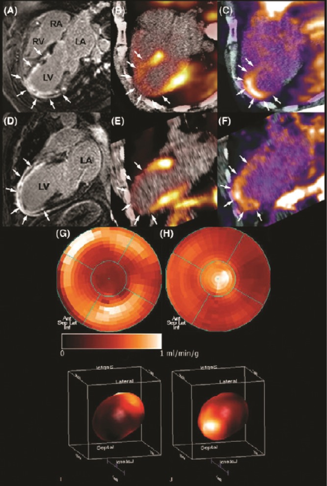
Figure 4.

In vivo molecular imaging of angiogenesis in a patient after acute myocardial infarction. Two weeks after acute myocardial infarction and percutaneous coronary intervention, a patient underwent cardiovascular magnetic resonance imaging (CMR) and positron emission tomography (PET)/computed tomography (CT). CMR gadolinium delayed contrast enhancement (DCE) was observed almost transmural in the anterior, anteriorseptal, anteriorlateral, and apical left ventricle wall (A, D). PET imaging with 13N-ammonia revealed impaired myocardial blood flow in correspondence with the regions of DCE (arrows; B, E). PET with the agent 18F arginine-glycine-aspartic acid peptide (18F-RGD), with affinity for the  integrins, demonstrated focal signal areas in the infarcted area (C, F). This signal may reflect angiogenesis within the healing area (arrows). Polar maps of myocardial blood flow assessed by PET with 13N-ammonia (G, I) show severely reduced blood flow in the distal left anterior descending coronary artery perfusion region. Co-localized 18F-RGD signal corresponded to the regions of reduced blood flow (H, J), demonstrating the extent of the
  integrin expression in the infarcted area. Reprinted with permission from Makowski et al.107
