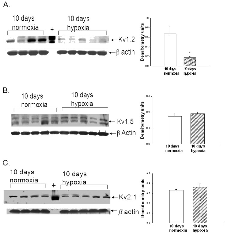
Figure 5.
A, Immunoblot results and corresponding densitometry for Kv1.2 α subunit protein relative to β actin in pulmonary arteries from normoxic and hypoxic piglets of the 10-day exposure group. The plus sign denotes positive control (rat brain). An asterisk indicates a significant different from normoxic piglets ( ). B, Immunoblot results and corresponding densitometry for Kv1.5 α subunit protein relative to β actin in pulmonary arteries from normoxic and hypoxic piglets of the 10-day exposure group. C, Immunoblot results and corresponding densitometry for Kv2.1 α subunit protein relative to β actin in pulmonary arteries from normoxic and hypoxic piglets of the 10-day exposure group. The plus sign denotes positive control (rat brain). In all panels, all values are mean ± SEM.

