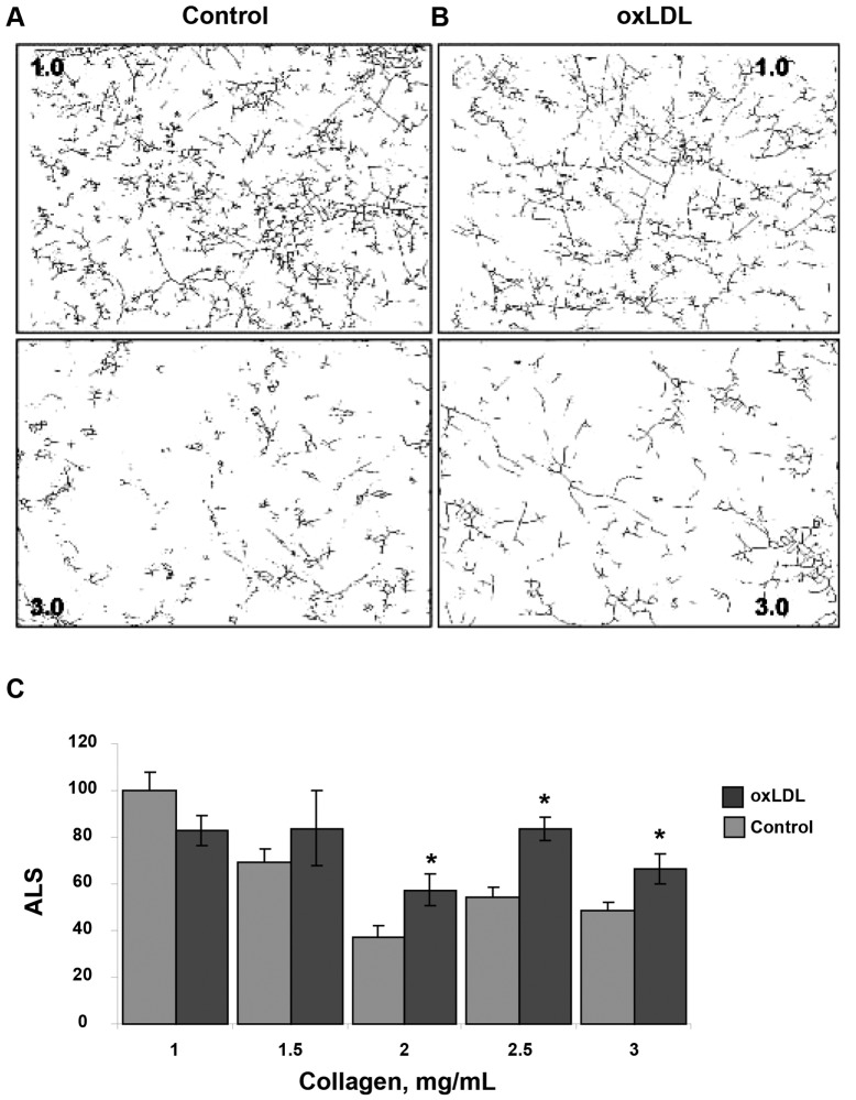
Figure 2.
Effect of oxidized low-density lipoprotein (oxLDL) on endothelial network formation in gels of different collagen concentrations. A, B, Representative skeletonized images of endothelial network shown in 1.0- and 3.0-mg/mL collagen gels for control cells (A) and for cell preexposed to 10 μg/mL oxLDL (B), demonstrating that the network is significantly suppressed in 3.0-mg/mL collagen gels as compared to 1.0-mg/mL gels. The upper panels show the images of the network in soft gels (1.0 mg/mL), and the lower panels show the images in stiff gels (3.0 mg/mL). C, Average length of skeletonized image (ALS), an index of cell elongation and network connectivity, as a function of collagen gel concentration for control and oxLDL-treated cells. All values are normalized to the ALS index of control cells seeded in gels of the lowest stiffness (1 mg/mL collagen) in the same experiment. The data show mean ± standard error of the mean; an asterisk shows significance between control and oxLDL-treated cells ( ).
).

