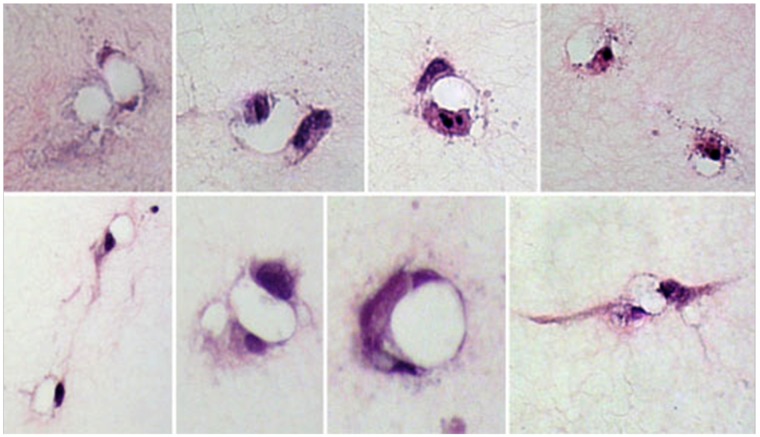
Figure 3.
Representative images of endothelial cell lumens (matrix-free regions surrounded by cellular regions) formed in the collagen gels demonstrates the various shapes and sizes of lumens formed, with cellular regions being made up of 1 or more endothelial cells and their extensions. The images were taken at 200×.

