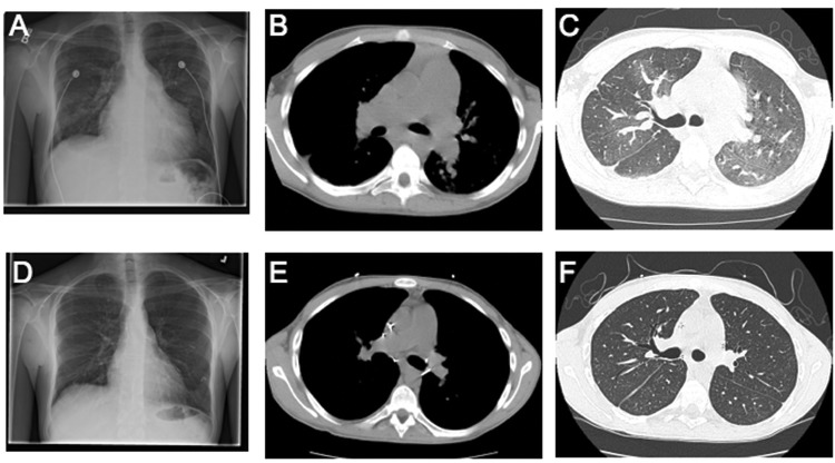
Figure 3.
Comparison of pre- and posttransplant radiologic changes. Pretransplant chest radiograph (A) and CT scans (B, C) demonstrate an enlarged right ventricle and enlarged pulmonary arteries. In addition, patchy mosaicism is seen in the parenchyma (C). These findings are resolved with transplant, as seen in the virtually normal posttransplant chest radiograph (D) and CT scan (E, F).

