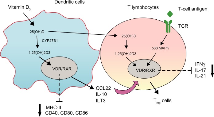
Figure 2.

Schematic representation of the primary mechanisms through which vitamin D-regulated dendritic cells (DCs) and T-lymphocyte function.
Notes: vitamin D precursors can be further processed to their active metabolite, 1,25(OH)2D3, in DCs and T lymphocytes. In DCs, 1,25(OH)2D3 binds to the vitamin D receptor–retinoid X receptor (VDR/RXR) complex in the nucleus, leading to a tolerogenic DC phenotype, characterized by decreased expression of major histocompatibility complex (MHC)-II, CD40, CD80, CD86, enhanced expression of immunoglobulin-like transcript (ILT)-3, and increased secretion of interleukin (IL)-10 and CCL22, which results in the induction of T-regulatory (Treg) cells. The 1,25(OH)2D3 signaling in T-cells is dependent on the stimulation of T-cell antigen-receptor (TCR) signaling. VDR expression can be induced by TCR signaling via the alternative p38 MAPK pathway. 1,25(OH)2D3 binds to VDR, leading to inhibition of proinflammatory cytokine expression, including interferon (IFN)-γ, IL-17, and IL-21, and promotion of the development of Treg cells.
Abbreviations: CCL22, chemokine (C-C motif) ligand 22; MAPK, mitogen-activated protein kinase.
