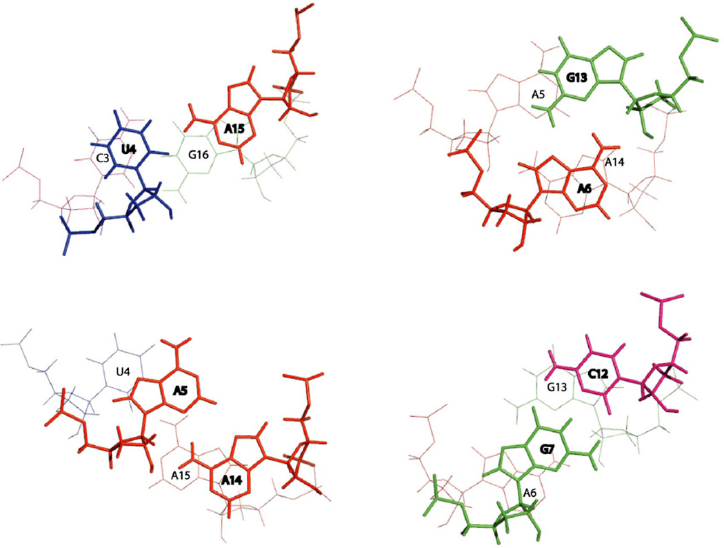
Figure 8.
Stacking patterns of the residues in the loop generated from the average of the 20 lowest free energy structures using 3DNA (97). Adenines are colored red, cytosines are pink, guanines are green and uracils are blue. The base pair closer to the viewer is in bold.

