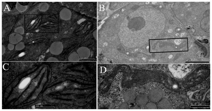
Figure 3. Transmission electron microscopy analyses of chloroplast biogenesis in the ftshi4 mutant embryos.
A, Wild-type torpedo embryo from a heterozygous ftshi4-1 plant with well-developed chloroplasts showing thylakoid membranes beginning to stack into grana. B, Mutant embryo from the same heterozygous ftshi4-1 silique with development-disrupted “plastids”. C, Enlargement of an above-described chloroplast indicated by an arrow.D, Enlargement of an above-described “plastid” indicated by an arrow.

