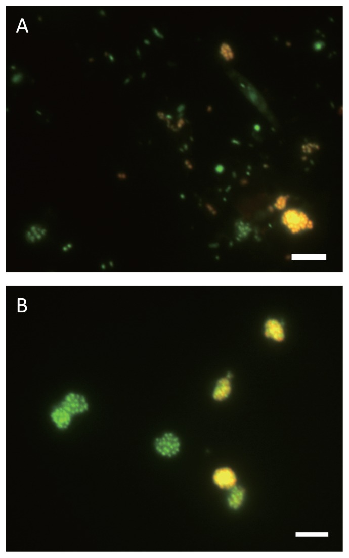
Fig. 1.
Fluorescence in situ hybridization (FISH) images of enrichment culture and separated micro-colonies. In situ hybridization was performed with Cy3-labeled probe Ntspa1151, specific for the detection of Nitrospira sublineage II (red). Green cells were stained only with SYTOX green, which stains all the cells. Yellow signals resulted from binding of both the Cy3-labeled probe and SYTOX green to one cell. Both scale bars are 5 μm.
(A) Enrichment sample treated by sonication prior to sorting Nitrospira micro-colonies.
(B) Nitrospira micro-colonies obtained from P6.

