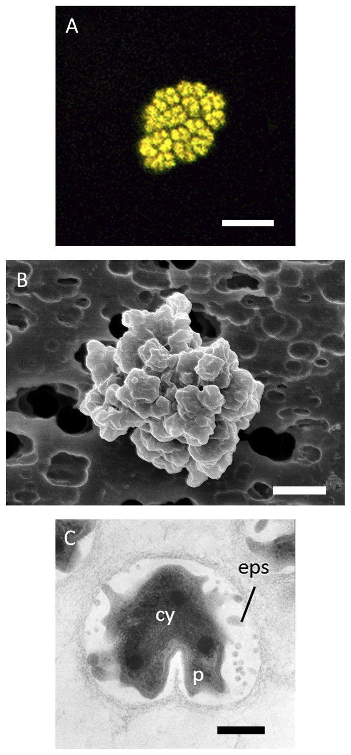
Fig. 4.
Morphology of Nitrospira japonica J1 isolated from activated sludge.
(A) In situ hybridization was performed with Cy3-labeled probe Ntspa1151, specific for Nitrospira sublineage II (red) and mixed FITC-labeled probes of EUB338, EUB338II, and EUB338III, for the detection of all bacteria (green). Nitrospira japonica appeared yellow due to binding with both Cy3-labeled and FITC-labeled probes. Scale bar is 5 μm.
(B) Scanning electron microscopic image of a micro-colony. Scale bar is 1 μm.
(C) Ultrathin section of a micro-colony revealing the wide periplasmic space and extracellular polymeric substances. cy = cytoplasm, p = periplasm, eps = extracellular polymeric substances. Scale bar is 200 nm.

