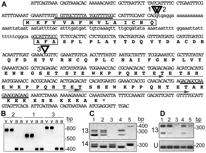
Figure 3. vH13 candidate gene 13 structure and expression in H13-virulent and avirulent strains.
(A) H13-avirulent genomic DNA sequence of vH13 candidate-13 showing exons (capital letters), intron (lower case letters), PCR primer-targeted sites (bold underlining), the positions of virulence-associated insertions (triangles 1, 2 and 3) and the predicted amino acid sequence (bold letters). The predicted signal peptide is boxed and the three imperfect direct repeats are underlined with arrows. (B) Candidate-13 fragments amplified using genomic DNA template extracted from H13-virulent (v) and H13-avirulent (a) individuals. H13-virulence associated sequences correspond to the insertions (1, 2 and 3) shown in panel A. For an explanation of the band lengths, see Figure S3. (C) Candidate-13 (13) and candidate-14 (14) transcripts amplified using total RNA extracted from pools of first-instar larvae (KS-GP, lane 1; IN-L, lane 2; vH13, lane 3 and IN-vH9, lane 4). Only candidate-14 sequence was amplified using the RNA extracted from the pool of H13-virulent first-instar (vH13, lane 3). Genomic DNA extracted from a single KS-GP larva was amplified as a control (lane 5). (D) Amplification of candidate-13 (13) and HF-ubiquitin (U) gene sequences using total RNA extracted from pools of H13-avirulent first-instars (lane 1), second-instars (lane 2), third-instars (lane 3), first-instar salivary glands (lane 4), and the carcases of first-instar larvae after salivary gland removal (lane 5).

