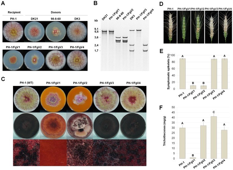
Figure 1. Effects of Fusarium mycovirus infections on F. graminearum strain PH-1.
PH-1/FgV1, PH-1/FgV2, PH-1/FgV3, and PH-1/FgV4 indicate the PH-1 strain infected with FgV1, FgV2, FgV3, and FgV4, respectively. (A) Colony morphology of Fusarium strains used in this study. PH-1 strains infected with FgV1–FgV4 were obtained by protoplast fusion. DK21 and 98-8-60 are FgV1 and FgV2-infected strains, respectively, whereas DK3 is an FgV3/4-coinfected strain. Cultures were photographed after 5 days on PDA. (B) Viral dsRNAs were extracted from mycelia using CF-11 cellulose chromatography. The presence of dsRNAs was confirmed by 5% polyacrylamide gel; the gels were stained with EtBr and visualized with a UV-transilluminator. (C) Sexual development of each strain. The strains were incubated on carrot agar medium for 7 days (upper row) and then treated with a Tween-60 solution to induce sexual reproduction (middle row). Cultures were examined for perithecia after 7 days under UV light. Scale bar = 200 µm. (D) Fusarium head blight symptoms caused by PH-1 (WT) or mycovirus-infected strains PH-1/FgV1–4. (E) Disease severity was evaluated 14 days after inoculation. (F) Trichothecene production. Conidial suspensions were grown in minimal medium containing 5 mM agmatine. After 7 days, trichothecenes in the culture filtrates were quantified by GC-MS. For (E) and (F), data were analyzed by the General Lineal Model (GLM) using IBM SPSS statistics 20 for Windows software. Error bars indicate standard deviations. Different letters above the bars indicate significant differences at p = 0.05.

