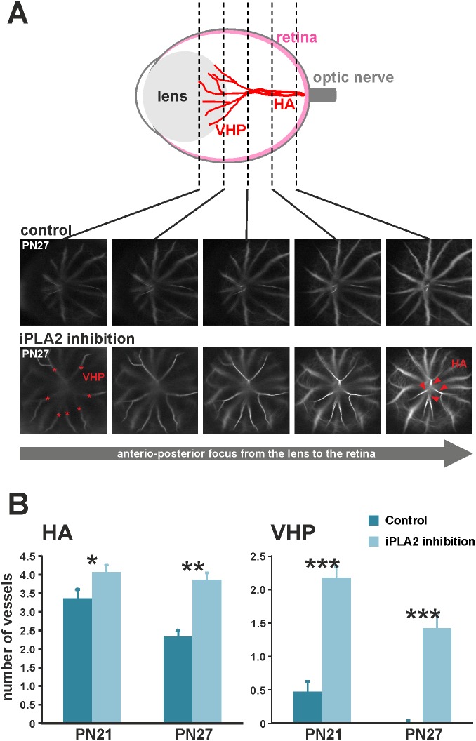
Figure 7. Defects in hyaloid vasculature regression in mice with iPLA2 inhibition.
A. Quantification of hyaloid arteries (HA) and vasa hyaloidea propria (VHP) vessels on depth-scan images from confocal cSLO angiography. VHP vessels (stars) were visualized and quantified at the level of the posterior lens, whereas HAs (arrowheads) were counted in the posterior eye in control and iPLA2-inhibited animals (n = 11 per group). B. Quantitative evaluation of hyaloid arteries (HA) and vasa hyaloidea propria (VHP) vessels in control and iPLA2-inhibited mice at PN21 and PN27. The numbers of HAs and VHPs were significantly higher in iPLA2-inhibited mice at PN21 and PN27 when compared to controls, thus confirming that the control of hyaloid vessel regression by Pls involves the iPLA2 enzyme. *: Statistically significant difference when compared to control group (Kruskal-Wallis test, P<0.05); **: statistically significant difference when compared to control group (Kruskal-Wallis test, P<0.01); ***: statistically significant difference when compared to control group (Kruskal-Wallis test, P<0.001).

