Fig. 3.

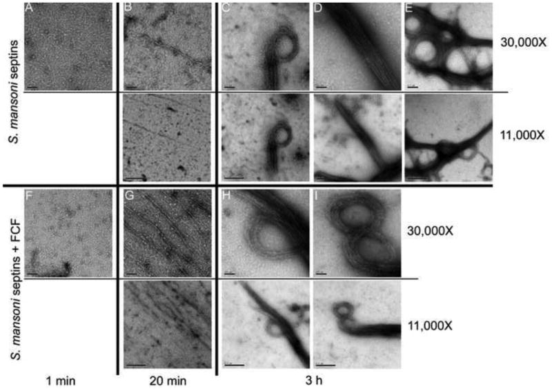
Schistosoma mansoni septin complexes assemble into filaments in vitro. Transmission electron micrographs of negatively stained septin complexes after dialysis against low salt solution with DMSO (A - E) or Forchlorfenuron (FCF) (F - I) for: 1 min (A, F), 20 min (B, G) or 3 h (C - E, H, I). The diversity of higher-order structures formed by S. mansoni septins is especially evident after 3 h of dialysis. Nominal magnification for each panel is shown on the right.
