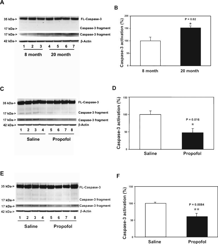
Fig. 3.
Propofol attenuates the aging-associated caspase-3 activation in cortex and hippocampus of WT mice. A) There is a greater caspase-3 activation in the cortex of 20 month-old WT mice (lanes 4-7) as compared to 8 month-old WT mice (lanes 1-3). There is no significant difference in amounts of β-Actin between the two groups. B) Quantification of the western blot shows that there is a greater caspase-3 activation in the cortex of 20 month-old WT mice (black bar) as compared to that of 8 month-old mice (white bar). C) Propofol (lanes 5-8) decreases caspase-3 activation as compared to saline (lanes 1-4) in the cortex of 20 month-old WT mice. There is no significant difference in amounts of β-Actin in saline- or propofol-treated mice. D) Quantification of the western blot shows that propofol (black bar) decreases the caspase-3 activation as compared to saline (white bar) in the cortex of the 20 month-old WT mice. E) Propofol (lanes 5-8) decreases caspase-3 activation as compared to saline (lanes 1-4) in the hippocampus of 20 month-old WT mice. There is no significant difference in amounts of β-Actin in saline- or propofol-treated mice. F. Quantification of the western blot shows that propofol (black bar) decreases the caspase-3 activation as compared to saline (white bar) in the hippocampus of 20 month-old WT mice. FL, full length; WT, wild-type. n = 6.

