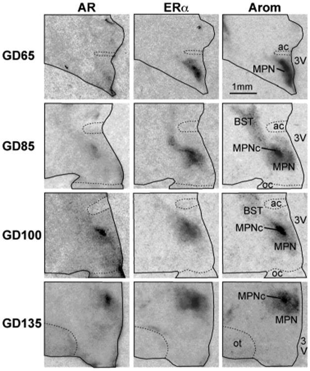
Figure 3.

Digitized autoradiographic film images of coronal sections through the right half of the fetal lamb preoptic region showing the pattern of expression for ERα and AR mRNAs at gestational days (GD): 65, 85, 100 and 135. Note that the dark signal for aromatase mRNA in the central area of the medial preoptic n. (MPNc) represents the nascent oSDN. All sections are from males, no significant differences were found in the patterns of the signal in females. Sections were cut at 20 μm thickness except for GD135 which was cut at 40 μm. All images were enlarged 380× (see bar for scale). Abbreviations: ac, anterior commissure; BST, bed n. of the stria terminalis; MPN, medial preoptic n.; oc, optic chiasm; ot, optic tract; 3V, third ventricle. Brightness and contrast were adjusted to optimize images.
