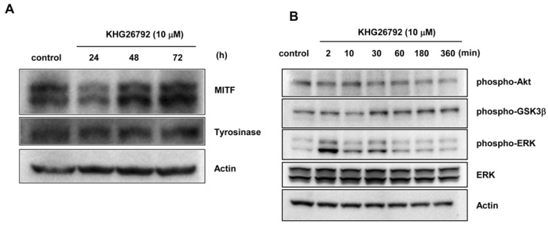
Fig. 3.
Effects of KHG26792 on melanogenic proteins and related signaling pathways. (A) Mel-Ab cells were cultured with KHG26792 (10 µM) for 24, 48, and 72 h. Western blot analysis was conducted with antibodies specific to MITF and tyrosinase. (B) After 24 h of serum-starvation, Mel-Ab cells were treated with 10 µM KHG26792 for 0~360 min. Cell lysates were assayed by Western blot analysis using antibodies against phospho-Akt, phospho-GSK3β, and phospho-ERK. Equal protein loading was confirmed using an anti-actin antibody

