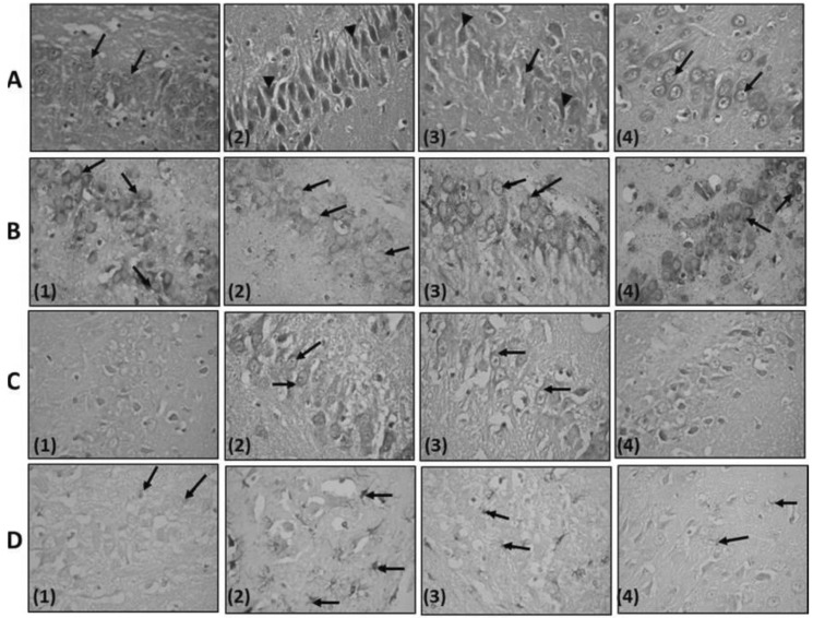
Fig. 7.
(rows A-D): A photomicrograph of CA3 region of the hippocampus: Row A, - Stained with H&E, ×540; (1) Control rat: notice that the pyramidal layer is formed of closely packed pyramidal cells with vesicular nuclei (↑). (2) Pentylenetetrazole (PTZ) treated rat: showing the presence of many deeply stained cells having pyknotic nuclei and perineuronal spaces (▸) an apparent decrease in the number of the pyramidal cells. (3) PTZ-Lamotrigine (LTG) treated rat: Pyramidal cells having vesicular nuclei and basophilic cytoplasm (↑). Few shrunken, darkly stained neurons are seen (▸) within the pyramidal layer. (4) LTG treated rat: nearly similar to control group (↑). Row B, Stained with Toluidine blue ×540: (1) Control rat: cytoplasm of the pyramidal cells appears studded with Nissl granules (↑). (2) PTZ treated rat: an apparent decrease in the Nissl granules of the pyramidal cells (↑). (3) LTG-PTZ treated rat: an apparent increase in the Nissl granules content of the pyramidal cells (↑) compared to that of PTZ treated rats. (4) LTG treated rat: picture nearly similar to control group. Row C, stained with Caspase-3 ×540; (1) Control rat: negative caspse-3 immuno- reaction in pyramidal cells. (2) PTZ treated rat: positive brownish caspase-3 immuno- reaction in pyramidal cells (↑). (3) PTZ-LTG treated rat: positive brownish caspase-3 immuno- reaction in few pyramidal cells (↑) when compared to PTZ group. (4): LTG treated rat: negative caspase-3 immuno- reaction in pyramidal cells. Row D, stained with Glial fibrillary acidic protein (GFAP) ×540. (1) Control rat: positive reaction for GFAP immuno-staining in few astrocytes (↑). (2) PTZ treated rat: an apparent increase in astrocytes (↑) size with longer cytoplasmic processes, number and GFAP immunoreactivity compared to control rats. (3) PTZ-LTG treated rat: an apparent decrease in the number of GFAP immunoreactive astrocytes (↑) compared to PTZ group. (4) LTG treated rat: few GFAP immunoreactive astrocytes (↑).

