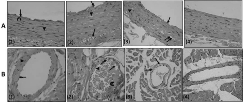
Fig. 8.
(row A, B): Row A, A photomicrograph of a transverse section in a rat thoracic aorta (H&E, ×540); (1) Control rat: the tunica intima lined with endothelial cells (↑), tunica media (M) is relatively thick and formed mainly of wavy acidophilic elastic laminae ( ) and smooth muscle cells (▲). Notice tunica adventitia is thin. (2) PTZ treated rat: clumps of cells with darkly stained nuclei (↑) attached to the endothelium of tunica intima which is apparently irregular. Few of the smooth muscle cells of tunica media show foamy acidophilic cytoplasm with karyolitic nucleus (▲) or clearly vacuolated cytoplasm with peripheral flat pyknotic nucleus (↑↑). (3) PTZ-LTG treated rat: apparently regular tunica intima (↑). Most of the smooth muscle cells are nearly similar to control group, others show foamy acidophilic cytoplasm (↑↑) and few number shows vacuolated cytoplasm with deeply stained nucleus (▲). (4) LTG treated rat: tunicae of the aorta are nearly similar to the control group. Row B, A photomicrograph of a section a coronary artery of the heart (H&E, ×540); (1) Control rat: an intact endothelial lining (↑). The tunica media (M) is formed of smooth muscle fibres with rod shaped vesicular nuclei (▲). The adventitia (A) is formed of loose connective tissue. (2) PTZ treated rat: multiple vacuoles in the tunica intima and tunica media (↑). Notice adherence of blood elements to the endothelium (▲). Exudates are also seen (
 ). (3) PTZ-LTG treated rat: few vacuolated cells are seen in tunica media (↑). (4) LTG treated rat: nearly intact tunicae.

