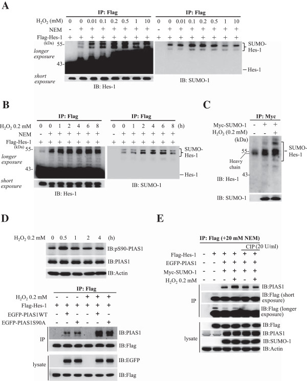
Figure 2.

H2O2 enhances the SUMOylation of Hes-1. (A) Flag-Hes-1WT plasmid was transfected to HEK293T cells with the addition of different concentrations of H2O2 and the cell lysate was immunoprecipitated with anti-Flag antibody and immunoblotted with anti-Hes-1 (left) or anti-SUMO-1 (right) antibody. (B) Flag-Hes-1WT plasmid was transfected to HEK293T cells with the addition of 0.2 mM H2O2 for different time periods. The cell lysate was immunoprecipitated with anti-Flag antibody and immunoblotted with anti-Hes-1 (left) or anti-SUMO-1 (right) antibody. (C) Myc-SUMO-1 plasmid was transfected to HEK293T cells with the addition of 0.2 mM H2O2 for 4 h. The cell lysate was immunoprecipitated with anti-Myc antibody and immunoblotted with anti-Hes-1 and anti-SUMO-1 antibody. (D) H2O2 (0.2 mM) was added to HEK293T cells for different time periods and the cell lysate was subject to western blot against pSer-90 PIAS1 and PIAS1 (upper panel). Flag-Hes-1WT plasmid was transfected to HEK293T cells with co-transfection of EGFP-PIAS1WT or EGFP-PIAS1S90A plasmid and with the addition of H2O2 for 4 h and the cell lysate was immunoprecipitated with anti-Flag antibody and immunoblotted with antibodies against PIAS1 and Flag. Western blot against EGFP and Flag for cell lysates only was used as a control for transfection and expression (lower panel). (E) Flag-Hes-1WT plasmid was co-transfected with EGFP-PIAS1 and Myc-SUMO-1 plasmids, and H2O2 was added to some of these groups for 4 h. The phosphatase inhibitor CIP (20 U/ml) was also added to some of these groups. The cell lysates were immunoprecipitated with anti-Flag antibody and immunoblotted with anti-PIAS1 antibody. Western blot against Flag, PIAS1 and SUMO-1 for cell lysates only was used as loading controls. The sumoylated Hes-1 bands are shown in the bracket. Each experiment was performed twice.
