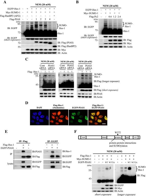
Figure 3.

PIAS1 is associated with Hes-1 and PIAS1 enhances the SUMOylation of Hes-1 in HEK293T cell. (A) Transfection of various plasmids to HEK293T cells followed by western blots against EGFP and various tags. (B) EGFP-Hes-1, Myc-SUMO-1 and different doses of Flag-Pc2 plasmids were transfected to HEK293T cells and western blots against EGFP and different tags are shown. (C) siRNA against PIAS1, PIAS2 and PIAS3 was transfected to HEK293T cells and western blots against Flag and PIAS proteins (PIAS1, PIAS2 and PIAS3) are shown. (D) Flag-Hes-1WT and EGFP-PIAS1WT plasmids were co-transfected to HEK293T cells and confocol image showing the sub-cellular localization of Hes-1 (red) and PIAS1 (green). The nuclei were visualized with DAPI staining (blue). Scale bar equals 10 μm. (E) Flag-Hes-1WT and EGFP-PIAS1WT plasmids were transfected to HEK293T cells followed by immunoprecipitation and western blot showing Hes-1 association with PIAS1 (left panel) and vice versa (right panel). (F) Various combinations of Flag-Hes-1, Myc-SUMO-1, EGFP-PIAS1WT or EGFP-PIAS1W372A plasmid were co-transfected to HEK293T cells and the cell lysate was subject to western blot against various tags. The sumoylated Hes-1 bands are shown in the bracket. Each experiment was performed twice.
