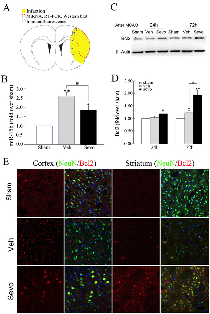
Figure 2. Sevoflurane preconditioning suppresses the ischemic induction miR-15b and allows Bcl-2 protein expression.
(A) The dashed red circle indicates the region of infarct in the cortex from which tissue was examined for the microarray data and RT-PCR, whereas the blue boxes indicate the regions in the cortex and striatum used for Bcl2 expression experiments. (B) Validation of microarray data and effects of sevoflurane preconditioning using quantitative RT-PCR. The level of miR-15b was up-regulated in vehicle relative to sham-operated rats at 72h after MCAO. Real-time PCR analysis of miR-15b in vehicle and sevoflurane preconditioning groups was carried out (n=6/group). Triplicate assays were performed for each RNA sample and the relative amount of each microRNA was normalized to U6 RNA. (C) Representative Western blot of Bcl-2 in ipsilateral cortex at 24 or 72 hours after reperfusion. (D) Semi-quantitative analysis of Bcl-2. All data are mean± SE. n=6/group. *P<0.05, **P<0.01 compared to sham-operated rats; #P<0.05 compared to vehicle rats. (E) Expression of Bcl-2 protein in neurons preconditioned with sevoflurane increases at 72 h after ischemia. Representative images of NeuN (green) and Bcl2 (red) immuno-fluorescence double-labeling in a peri-infarct region in ipsilateral (Ipsi) cerebral cortex (Ctx) and striatum (Str) in sham-operated, vehicle- and sevoflurane preconditioning rats at 72 hours after reperfusion. Scale bar =100 µm.

