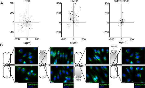
Figure 1.

BMP2 induces chemotaxis of multipotent mesenchymal C2C12 mouse myoblasts. (A) Trajectories of multipotent mouse mesenchymal C2C12 cells migrating in a 2D chemotaxis chamber over period of 16 hours exposed to a linear BMP2 gradient compared to non-stimulated control or in the presence of the PI3K p110α selective inhibitor PI103 (8 nM). The gradient was produced by application of BMP2 to the upper reservoir. It was allowed to generate a linear concentration profile with a maximum concentration of approximately 10 nM reaching the cells on the edge of the observation area as described by the manufacturer. (B) Syntaxin 6 and DAPI stainings of C2C12 cells after BMP2-induced chemotaxis compared to non-stimulated control or PI103 [8 nM] pre-treatment. The location of the depicted cells within the chemotaxis chamber is indicated. Scale bar represents 20 μm. (2D) two dimensional; (BMP2) Bone Morphogenetic Protein 2; (PI3K) Phosphoinositide 3-kinase; (p110α) Class I PI3K catalytic subunit alpha; (DAPI) 4',6-diamidino-2-phenylindole.
