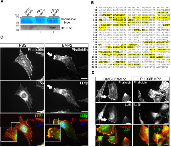
Figure 6.

The PIP3-binding protein LL5β localises to BMP2-induced cortical actin-rich lamellipodia. (A) Upper panel shows colloidal Coomassie Blue staining of protein precipitates gained by precipitation of PIP2-, PIP3-coated and control beads from C2C12 cell lysates. Lower panel shows LL5β detection (approximately 160 kDa) by western blot. LL5β binds to PIP3 (lane 3) but not PIP2 or control beads (lanes 1 and 2). (B) LL5β-specific peptides (marked in yellow) as identified by mass spectrometry upon precipitation of PIP3-coated beads from C2C12 cell lysates. (C) Immunocytochemical stainings of endogenous LL5β and actin in C2C12 cells upon 45 minutes’ stimulation with 10 nM BMP2. Arrow indicates co-localisation of LL5β with cortical actin in BMP2-induced cell protrusions at the C2C12 cell leading edge (magnified region of interest). (D) PI103 pre-treatment blocks BMP2-induced co-localisation of LL5β with cortical actin. C2C12 cells were stimulated with 10 nM BMP2 for 45 minutes in the presence of DMSO or 8 nM PI103 respectively. The magnified region of interest depicts the co-localisation of LL5β with cortical actin. Scale bars represent 20 μm.
