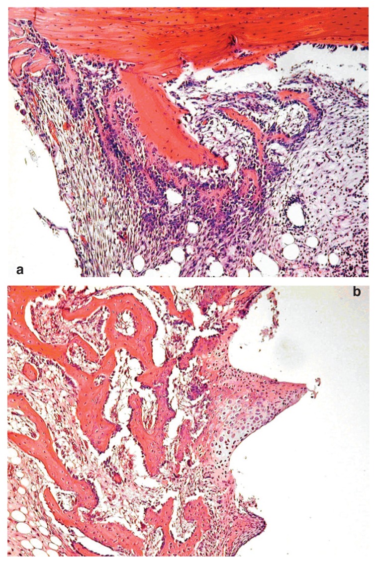
Figure 1.
Histological analysis of the implant site treated with PDGF and IGF-1. 1a) At 4th day, the implant sites treated with PRP or PDGF/IGF1 showed abundant cap-like deposition of fibroblastic tissue around the implants; this fibrous tissue was composed of spindle cells with elongated or oval nuclei and numerous mitotic figures. 1b) At the 12th day, a substantial amount of well formed, significantly more abundant woven bone in subjects treated with PDGF/IGF1 than in animals treated with PRP and controls, was found.

