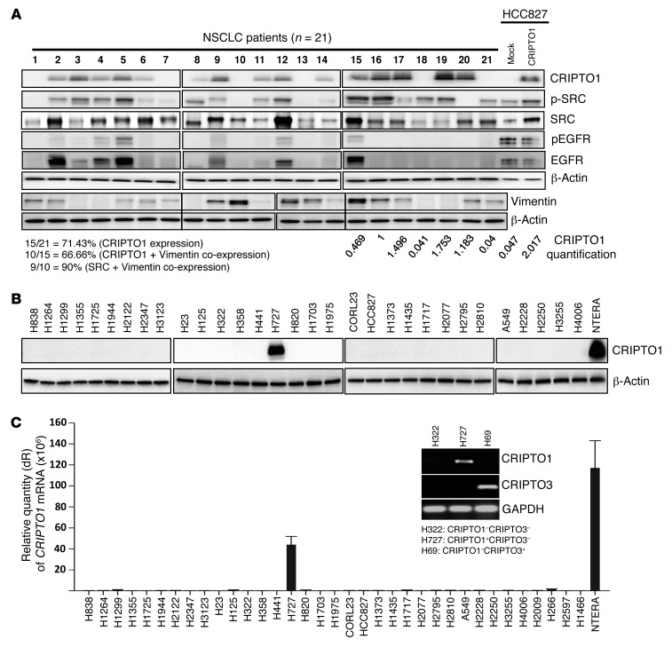
Figure 1. CRIPTO1 expression in NSCLC tumors and cell lines.
(A) Western blot analysis of CRIPTO1, total SRC, phosphorylated SRC, pEGFR, EGFR, and Vimentin in 21 NSCLC patient samples. Relative expression of endogenous CRIPTO1 in tumor cells and exogenous CRIPTO1 in HCC827 cells were shown after normalization against β-actin (lanes 15–21, HCC827 mock, and HCC827/CRIPTO1). Note that lanes 1–14 were not used for comparison, as they were derived from different blots, and that the lanes on 2 sides of the thin black lines were run on the same gel but were noncontiguous. (B) CRIPTO1 expression in 31 NSCLC cell lines by Western blot. (C) CRIPTO1 mRNA expression in 35 NSCLC cell lines by RT-PCR. CRIPTO1 primers could only amplify CRIPTO1 in CRIPTO1-positive/CRIPTO3-negative (H727) cells, but not CRIPTO3 in CRIPTO3-positive/CRIPTO1-negative (H69) cells (inset).

